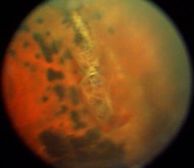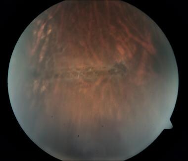

Most of the current approaches to clinical management are based on indirect ophthalmoscopic interpretation. Asymptomatic retinal breaks may lead to chronic inferior retinal detachment and may occasionally be difficult to distinguish from retinoschisis. Findings such as lattice degeneration, white without pressure, vitreoretinal traction, and posterior vitreous detachment- (PVD-) associated retinal breaks are common reasons for the need to treat with retinopexy in order to prevent retinal detachment. Peripheral vitreoretinal interface abnormalities span a range of entities from incidental ophthalmoscopic findings to retinal detachment. This imaging modality is useful in the clinical management of suspected retinal breaks identified with indirect ophthalmoscopy. Peripheral SD-OCT is a reliable and useful technique to examine the structural features of vitreoretinal interface abnormalities in vivo. Two cases of previously diagnosed operculated holes were found on SD-OCT to be partial-thickness operculated breaks or focal operculated schisis. Decision to treat was altered following SD-OCT in 5% of the patients. Mean age was 41 ± 22 years, and mean follow-up was 14 ± 1.6 months. Acceptable image quality for inclusion was obtained in 39/43 (91%) patients. Laser retinopexy was performed to surround all retinal breaks containing a full-thickness component via SD-OCT.

SD-OCT was evaluated for image quality and structural findings. A prospective imaging analysis of 43 patients with peripheral vitreoretinal interface abnormalities seen on binocular indirect examination with scleral indentation was done. Synergy Eye Care is well equipped and its doctors are well experienced in treating this disease using required procedures and /or surgeries with good results.ĭisclaimer: Information published here is for educational purposes only and is not intended to replace medical advice.The objective of this study is to describe the clinical utility and morphologic characteristics of peripheral vitreoretinal interface abnormalities with spectral domain optical coherence tomography (SD-OCT).

Therefore the patients need to maintain regular retinal checkups in the future. However one must understand that despite the treatment, there is still a small risk of retinal detachments or appearance of new lesions. These Delimiting Laser or Cryotherapy are very safe with practically no significant complications. This is generally recommended when the other eye has had a retinal detachment, the lattice appears more dangerous, presence of retinal tear, strong family history of retinal detachment, etc.Īre there any side-effects of the Laser Treatment or Cryotherapy?


 0 kommentar(er)
